Illustrations in Medicine

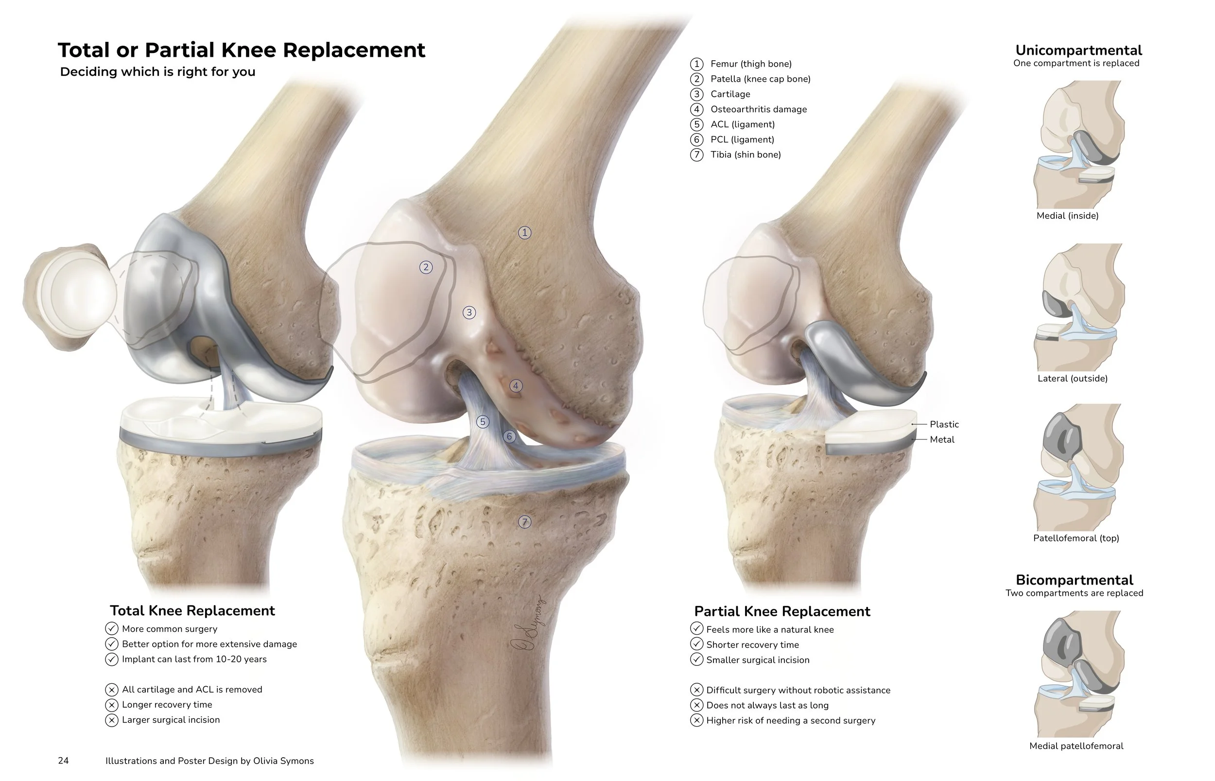
Patient education is a crucial step in healthcare decision making. Scientific illustrations and infographics are resources that transcend language barriers and facilitate better communication between patients and healthcare providers.

Illustrations can also play a vital role in teaching and sharing knowledge. They help visualize new medical procedures, educate student doctors, and obtain grant funding for research projects.

Here are some examples of illustrations made for the medical field.

Detailed diagram comparing total and partial knee replacement, including labeled parts of the knee joint such as femur, patella, cartilage, osteoarthritis damage, ACL and PCL ligaments, and tibia. It shows differences between unicompartmental and bicompartmental knee replacements, and illustrations of knee replacement options with descriptions of benefits and drawbacks.
Infographic on the role of robots in knee replacement surgery, detailing four steps: pre-operative plan, landmarking, ligament balancing, and bone preparation, with illustrations of robotic tools and knee anatomy.

See more:

Academia

Industry

Animal